
An Application of Collisions – Radiological Diagnostic Imaging
On hearing the word collisions, we may think about railway car accidents, movies, novels, other works of arts, ideologies, or expensive physical experiments on particles that can shape the models we use to describe the universe. But most people probably wouldn’t relate collisions to the two classical and widely used diagnostic imaging technologies: conventional X-ray and computed tomography (CT).
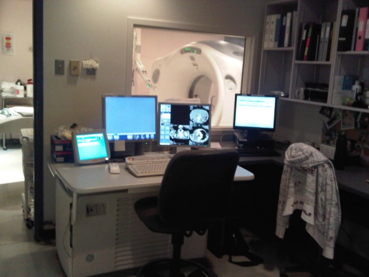 In the Diagnostic Imaging program at Dawson, we learn some of the science that makes these technologies possible, and it turns out that this kind of imaging involves collisions––three cascades of collisions. The actual order in which these collisions occur is the following:
- production of the X-rays
- interaction of the X-rays with the human body
- detection of the X-rays which allows the invisible radiological image, present in the space after the body, to be captured by the image receptor.
I believe that you, the readers, are more interested in the applications and not in the physics of the X-ray collisions. So, I will focus on the applications.
Both plain X-ray and CT are possible, first, because of the collisions between X-ray photons and the matter of the human body. More precisely, the collisions occur with different intensities when x-rays hit air, fat, water, soft tissue and bone. X-rays pass through air much more easily than they do through bone. This difference accounts for the fact that on a negative X-ray image, our lungs are black, whereas our bones are white, with some shades of gray in between. As well, the greyer the image, the less contrast and the more shades it contains.
An interesting fact is that in the ultrasound examinations, the bone tissue blocks the ultrasound and does not allow the structures behind the bone to be visualized, whereas in the magnetic resonance imaging (MRI), bone does not significantly attenuate the signal. For this reason, on a magnetic resonance image, the bones are black but the anatomy behind or around them is clearly visible. Because bone tissue produces no signal in the ultrasound examinations and in the magnetic resonance imaging, conventional X-ray and CT remain the modalities of choice for bone diagnostics. In other words, X-rays and CT are still, at the moment, irreplaceable technologies.
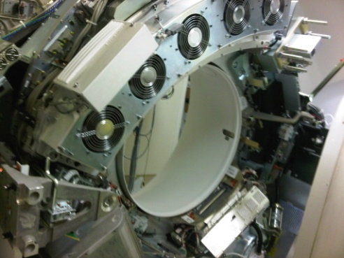
The discovery of X-rays, at the end of the XIX century, revolutionized medicine; doctors no longer needed to surgically open the body to see the morphological changes in it. Today, X-rays are still produced the way they were in 1895: in a vacuum through the collision of accelerated electrons with a target. The acceleration of the electrons is achieved by a very high voltage, and the target is the “anode” – the positive electrode which attracts the electrons. X-ray production is a very inefficient process. In fact, only one percent of the kinetic energy of the accelerated electrons accounts for the production of the X-rays, through what is called “breaking radiation” and “characteristic radiation”; the rest is heat. All x-ray tubes, used in diagnostic imaging, work on this collision principle and are coupled with highly efficient cooling systems. Thanks to the enormous heat capacity of the X-ray tubes (millions of heat units), continuous examinations can be done with no waiting time between the shoots.
Additionally, X-ray image detection itself is also achieved by collisions––collisions of X-rays with the image receptors, similarly to the way light collides with film. Unlike X-ray tubes which all use the same principle, image receptors exist in an enormous variety of realizations: from the old analog silver bromide films, the ionization xenon detectors, the scintillation detectors which convert X-ray to light photons, to the amorphous silicon, or the amorphous selenium flat panel receptors. My list does not include all the possibilities; however, the common feature of the image receptors is that they all use a certain type of collision to capture the image - the third, and the last cascade of collisions in the diagnostic imaging.
 The quality of the detected image would not withstand an artistic appraisal; it is just a collection of data corresponding to the internal anatomy for an in-depth technical analysis. Image processing and reconstruction algorithms are designed to provide the maximal possible information generated per study. For instance, the CT scan is always done in an “axial plane” (a plane perpendicular to the long axis of the body). On this axial plane the pathology is going to be visible, but the extent of this pathology will be better demonstrated on the “coronal” and “sagittal” reconstructions, which are performed along the long axes of the body. Additionally, the 3D reconstruction clearly demonstrates the volume of the pathological processes by showing whether the object of interest is as small as a needle top or big as a tennis ball, as well as by outlining the exact spatial configuration of the region of interest.
In the Diagnostic Imaging applications, the homogenously distributed X-rays from the source are directed toward the human body where two types of collisions in the tissues occur: partial absorption with scatter or total absorption, called “Compton and Photoelectric effects.” (The details and the differences between them are beyond the scope of this article.) As both collisions ionize some of the atoms of the body, the utmost goal of the conventional X-ray and computed tomography is to achieve sufficient diagnostic value by causing the minimum possible collisions in the human body. Image receptors which can efficiently cause and detect collisions--i.e. which have high detector quantum efficiency, contribute to the realization of this goal, thus helping to reduce the impact of these essential diagnostic tools on the body. The ultimate aim of the technologists, teachers and students of the Diagnostic Imaging Program at Dawson College is to work together to master these applications in order to “justify the trust the patient has in them.”
Comments
No comments posted yet.
You have to be registered and logged in in order to post comments!